Home
Dissection guides
3D Models
VR Lab
More
The dry skull
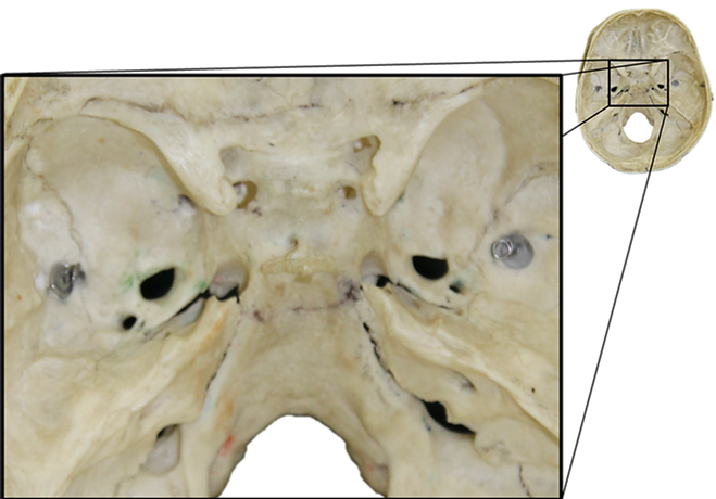
Crescent of foramina
The cranial nerves in the cadaver
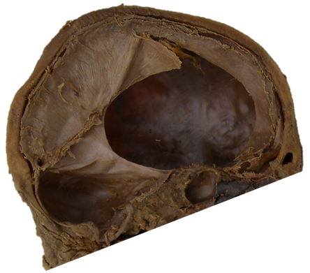
The endocranium in the cadaver
The dural sinuses