Cerebral white matter and basal nuclei The white matter of the hemispheres is located deep to the cortex and consists mostly of neuronal axons. These fibers are coated with a lipid-rich (fatty) substance called myelin that gives them the light color. The white matter fibers are organized in fasciculi (bundles) that connect the various areas of the nervous system.
There are three main types of fibers depending on the type of areas they connect:
Commissural fibers connect different areas between the two cerebral hemispheres.
Association fibers connect the different cortical areas within the same cerebral hemisphere.
Projection fibers connect the different cortical areas with subcortical structures.
We will start by examining the commissural fibers in a medial view of a midsagittal section of the hemisphere. Identify the different parts of the corpus callosum : the rostrum , genu , body and splenium . Find the remaining commissural systems: the anterior commissure is located between the lamina terminalis and the column of fornix , it connects the temporal lobes. The hippocampal commissure is located between the crus of fornix of each side and it connects the two hippocampi. Since it is very thin, it is harder to see it in a midsagittal section. The habenular commissure is located anterior to the pineal gland and it connects the two habenulae. The posterior commissure is located inferior and anterior to the pineal gland and it connects the superior colliculi .
The largest commissural structure is the corpus callosum . Notice that its medial part is shorter than the length of the hemisphere. The fibers that connect the anterior parts of the frontal lobes form the forceps minor and the fibers that connect the occipital lobes form the forceps major . The medial part of the corpus callosum is covered by a thin layer of gray matter, the indusium griseum . It is continuous with the dentate gyrus of the hippocampus. Its white matter is concentrated in two thin strips on each side, the medial longitudinal stria and the lateral longitudinal stria .
Superior view of corpus callosum
We will start the dissection by exposing the more superficial f ibers f irst and then, we will continue to the deeper f ibers and subcortical nuclei. We will begin by removing the cortex that covers the insula (island), the operculum (lid). Notice the thickness of the cortex (~2 mm) and the difference between it and the white matter.
Continue to remove the cortex around the insula . Notice the fibers that connect adjacent gyri, these are short association f ibers (U-fibers) . The other associative f ibers we will study are long-association f ibers. Find a long bundle of f ibers that bends around the insula and connects all of the lobes, the superior longitudinal fasciculus (SLF) . The fronto-temporal part of the SLF is also called arcuate fasciculus. Now, move to the limen of the insula and expose the uncinate fasciculus (hook) below it. It connects the anterior part of the frontal lobe with the anterior part of the temporal lobe. Find the inferior fronto-occipital fasciculus located above and behind the uncinate fasciculus, it connects the anterior inferior part of the frontal lobe and the occipital lobe. The inferior longitudinal fasciculus (ILF) connects the temporal lobe and the occipital lobe. It can be exposed by removing the gray matter from the lower surface of these lobes.
Next, proceed to the medial surface. By removing the cortex of the cingulate gyrus, expose a large structure of association fibers that connects all the lobes, the cingulum . Follow its fibers posteriorly where it passes through the isthmus (neck) and bends behind the splenium of corpus callosum . From there, it continues into the parahippocampal gyrus.
Anterior
and
medial
to
the
lentiform,
find
the
caudate
nucleus
(head)
and
posterior
and
medial
to
the
lentiform,
the
thalamus
.
between
these
three
structures,
notice
the
concave
shape
of
the
internal
capsule
and
three
of
its
five
parts:
The
anterior
limb
is
located
between
the
lentiform
and
caudate
and
it
contains
cortico-pontine
fibers
and
anterior
thalamic
radiation
f
ibers
that
connect
the
thalamus
and
frontal
lobe.
The
bent
central
part,
genu
,
is
located
medially
to
the
globus
pallidus
internus
and
it
contains
cortico
-nuclear
fibers.
The
posterior
limb
is
located
between
the
lentiform
and
thalamus
and
it
contains
cortico-spinal
fibers
and
f
ibers
that
connect
between
the
thalamus
and
somatosensory and association cortex.
The additional two parts of
internal capsule are the optic radiation (retrolenticular part) and the
auditory radiation (sublenticular part), which will be further exposed in
the following steps.
Also
identify
the
anterior
commissure
and
the
forceps
minor
and
forceps major
of the corpus callosum.
The
most
lateral
is
the
cortex
of
the
insula
and
its
white
matte
r
.
Medial
to
it,
find
the
thin
layer
of
white
matte
r
,
the
extreme
capsule
.
Medial
to
it,
identify
the
thin
layer
of
gray
matte
r
,
the
claustrum
(enclosed)
followed
by
another
thin
layer
of
white
matte
r
,
the
external
capsule
.
Continuing
mediall
y
,
find
the
lentiform
.
On
the
right,
its
three
parts
can
be
easily
distinguished
(from
lateral
to
medial):
the
putamen
(nutshell),
globus pallidus externus
and
globus pallidus internus
.
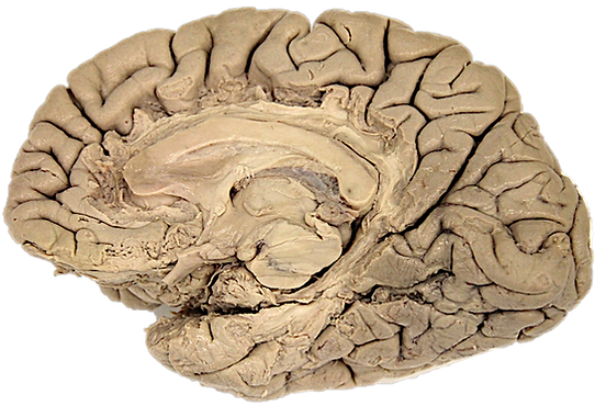
Horizontal section of the brain
For the next part of the dissection, we will use a horizontal section of the brain as a guide to the different structures we will discover as we remove the different layers from lateral to medial (note that the right hemisphere is cut lower than the left).
Return to the hemisphere you have started dissecting. Remove the gray matter of the insula and the thin layers of the extreme capsule, claustrum and external capsule. Continue removing the white matter until you reach a large lens-shaped nucleus, the lentiform . Finish exposing and defining the boundaries of the lentiform and remove the remaining association f ibers above it. Notice the fibers of the corona radiata that surround the lentiform, they connect between the cortex of the hemisphere and subcortical structures. Now that the insula is removed, the uncinate fasciculus and the inferior fronto-occipital fasciculus can be easily seen .
Gently separate the lentiform from the fibers that pass medially to it while keeping it intact. Once the lentiform is removed, you can see the internal capsule which is continues with the corona radiata. The fibers of the internal capsule that are posterior to the lentiform (retrolenticular part), constitute the optic radiation , they relay visual information between the lateral geniculate nucleus of the thalamus and the primary visual area that is located in the calcarine sulcus. The fibers of the internal capsule that pass under the lentiform (sublenticular part), are fibers of the auditory radiation , they relay auditory information between the medial geniculate nucleus of the thalamus and the primary auditory area that is located in the anterior transverse gyrus. Notice a bundle of fibers that passes under the lentiform and continues perpendicular to the orientation of the corona radiata, this is the anterior commissure . Next, Make a coronal section in the center of the lentiform and try to distinguish between the putamen and the globus pallidus.