Lateral ventricles and third ventricle
Ventricular system of the brain
The ventricular system is located deep in the brain and it contains cerebrospinal fluid (CSF). Its main parts are the two lateral ventricles, the third ventricle and the fourth ventricle.
Each lateral ventricle is connected to the third ventricle through an interventricular foramen (of Monro). The third and fourth ventricles are connected by the cerebral aqueduct (of Sylvius).
Three openings in the fourth ventricle connect the CSF of the ventricular system to the CSF of the subarachnoid space, one median aperture (of Magendie) and two lateral apertures (of Luschka). The fourth ventricle is also continuous with the central canal of the spinal cord.

Transverse fissure and tela choroidea
The transverse fissure separates between the inferior surface of the occipital lobes and the superior surface of the cerebellum. Through the transverse fissure, pia mater and arteries enter underneath the splenium to the roof of the third ventricle, where they form the tela choroidea of the third ventricle. The tela choroidea is continues with the choroid plexus of the lateral ventricles and they both produce the CSF. Similar structures exist in the fourth ventricle which we will study in another lab.


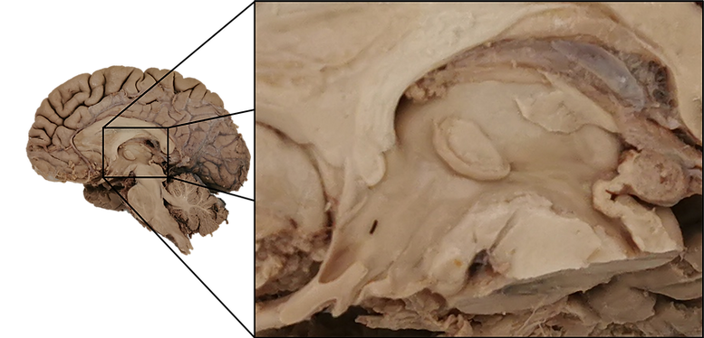
Third ventricle
Locate the oval shaped thalamus, its inferior boundary is the hypothalamic sulcus and inferior and anterior to it, is the hypothalamus. The third ventricle is the narrow space between the thalamus and hypothalamus of the two hemispheres. Along the border between the medial and dorsal surfaces of the thalamus, find a strip of white matter that contains fibers from the septal area to the habenular nuclei, the stria medullaris thalami. In the center of the thalamus, find the massa intermedia (interthalamic adhesion). At the roof of the third ventricle, find the tela choroidea and above it, the fornix. Between the column of fornix and the thalamus, find the interventricular foramen that connects the third ventricle with the lateral ventricle.
The tela choroidea is continuous with the choroid plexus of the lateral ventricle through the interventricular foramen and the choroid fissure (between the thalamus and fornix). The anterior boundary of the third ventricle is the lamina terminalis, between it and the column of fornix find the anterior commissure. Notice that of the third ventricle has four extensions (recesses). At the anterior part, the optic recess above the optic chiasm and the infundibular recess above the infundibulum.
At the posterior part, the suprapineal recess above the pineal gland and the pineal recess below it. At the bottom, the third ventricle is continuous with the cerebral aqueduct (of Sylvius) that connects it with the fourth ventricle.
Lateral ventricles
Each lateral ventricle consists of four parts: frontal horn, central part, occipital horn and temporal horn. For each part, we will learn the different structures that define the boundaries of the lateral ventricle.

Boundaries of the lateral ventricles
Frontal horn and central part of the lateral ventricles
Make a cut in central part of the corpus callosum and remove it:
This will expose the frontal horn and central part of the lateral ventricles.
The corpus callosum forms the roof and the septum pellucidum forms the medial wall that separates the two lateral ventricles. The frontal horn is anterior to the interventricular foramen and its floor is the head of caudate.
In the central part, the floor consists of the body of caudate and the thalamus (here it is covered by other structures). Between them, find the stria terminalis that contains fibers from the amygdala to the septal area and below it, the thalamostriate vein. In the central part of the lateral ventricle, notice how the choroid plexus starts at the interventricular foramen and continues posteriorly on top of the fornix.


Occipital horn of the lateral ventricle
Make a cut in the posterior part of the corona radiata: and reveal the occipital horn.
Notice that the choroid plexus continues into the temporal horn and does not reach the occipital horn. Also use a coronal section of the occipital horn and identify three bulges within it. The superior bulge is the bulb of occipital horn that is formed by the forceps major of the corpus callosum. The middle and largest bulge is the calcar avis, it is formed by the calcarine sulcus.
The Inferior bulge is the collateral eminence, it is formed by the collateral sulcus. The lateral wall is composed of fibers of the corpus callosum that continue downwards, the tapetum. Lateral to it, find the fibers of the optic radiation, that appears slightly darker because the cut was made perpendicular to the orientation of the fibers.


Temporal horn of the lateral ventricle
Make a cut in the white matter of the temporal lobe:
This will reveal the temporal horn. At the floor, find a large structure, the hippocampus (sea horse). It is a three-layer archicortex that is continuous with the six-layered neocortex of the parahippocampal gyrus.
Its anterior part is the pes hippocampus (paw). The white matter that covers the hippocampus is the alveus, along its medial line notice a separated fold of white matter, the fimbria (fringe) which is continuous with the fornix.
Make a coronal cut in the hippocampus and notice that its cortex folds on itself, the cornu ammonis (horns of Ammon). Along the medial line, below the fimbria, find a row of bulges, the dentate gyrus (teeth), it is continuous with the indusium griseum.
At the roof of the temporal horn, find the tail of caudate.
Also notice the choroid plexus.


Superior view of the third ventricle
Now that we have finished examining the parts of the lateral ventricles, return to the superior view of the ventricles. Make an incision in the columns of fornix and retract the two fornices backwards to reveal the third ventricle, the narrow space between the two thalami. Along their medial line, find the striae medullaris thalami. Gently separate the thalami and peer into the anterior part of the third ventricle. Find the two columns of fornix with the anterior commissure forming an "A" shape between them.