Endocranium and dural sinuses
The dry skull
Remove the calvarium of the skull and examine the interior of the cranium. The base of the endocranium is divided into three cranial fossae: anterior, middle and posterior. The anterior cranial fossa is separated from the middle cranial fossa by a bone ridge, the lesser wing of sphenoid bone. Identify it and follow it towards the midline, notice how it ends in two projections, the anterior clinoid processes. The middle cranial fossa is separated from the posterior cranial fossa by an elongated pyramid shape bone protrusion, the petrous part of the temporal bone.

Look at the center of the floor of the cranial cavity. Notice a saddle shaped raised structure, the sella turcica (Turkish saddle). It is defined by four processes: the anterior clinoid processes and the posterior clinoid processes. Between the two posterior clinoid processes identify the hypophyseal fossa, the site of the hypophysis (pituitary) gland. Anterior and medial to each anterior clinoid process find a circular opening leading to the orbital cavity, the optic canal.
The anterior cranial fossa contains the frontal lobe, in its medial part notice a perforated part of the bone, the cribriform plate through which the olfactory nerve fibres pass. Perpendicular to the cribriform plate, locate a comb shaped plate, the crista galli. The middle cranial fossa contains the temporal lobe. In its medial part, on either side of the sella turcica there is a series of foramina arranged in the shape of a crescent, the crescent of foramina.
Crescent of foramina
In the crescent of foramina identify the different openings from anterior to posterior. The superior orbital fissure through which cranial nerves: III, IV, V1 (ophthalmic branch) & VI pass. Next, is the foramen rotundum through which the nerve V2 (maxillary branch) passes. The next opening is the foramen ovale through which the nerve V3 (mandibular branch) passes. The most posterior small opening is the foramen spinosum through which the middle meningeal artery passes.
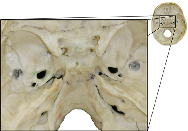
Near the anterior medial edge of the petrous, find the foramen lacerum. Next to it, lateral to the posterior clinoid process, identify the carotid canal that contains the internal carotid artery. Posterior to it, find the depression for trigeminal ganglion. From this region the trigeminal nerve (CN-V) splits into its three sensory branches: ophthalmic, maxillary and mandibular. Along the ridge of the petrous find a thin canal, the superior petrosal sinus.
The posterior cranial fossa contains the cerebellum. At its anterior part, in the posterior slope of the body of the petrous, find the internal acoustic meatus, through which cranial nerves VII and VIII pass. Posterior to it, there is a large hole that is formed in the space between the body of the petrous and the floor of the posterior cranial fossa, the jugular foramen. Through it, the internal jugular vein and the cranial nerves: IX, X & XI pass. At the bottom of the center of the posterior fossa find the foramen magnum. In the lateral part of it’s edges find the hypoglossal canal (anterior condylar) through which the cranial nerve XII passes.
At the midline of the posterior cranial fossa, find a thickening in the bone, the internal occipital protuberance. From this point try to identify the impression of the transverse sulcus, where the transverse sinus lies. Follow the canal laterally and notice how it winds down posterior to the petrous and ends in the jugular foramen. This tortuous section is the sigmoid sinus. Return to the internal occipital protuberance, notice a ridge that extends upward and continue sagittally along the entire length of the skull and reach the crista galli in the frontal bone, this is the superior sagittal sinus.
The endocranium in the cadaver
In the center of the endocranium, at the meeting point of the anterior cranial fossa and the middle cranial fossa, find the entry point of the optic nerve and posterior to it, the entry point of the internal carotid artery (the bone is removed on the left side). Notice the elevated structure of the sella turcica (Turkish saddle) and in its center the hypophyseal fossa, the seat of the hypophysis. In the posterior cranial fossa, find the foramen magnum through which the spinal cord continues.

The dural sinuses
Examine the dura mater in the endocranium. In the midline, identify a deep fold, the falx cerebri. It enters the longitudinal fissure between the two hemispheres. At its posterior margins, the falx cerebri splits sideways and forms the tentorium cerebelli that separates the occipital lobe from the cerebellum. Notice the anterior margins of the tentorium that surround the midbrain, the incisura tentorii.
Idetify two sinuses that run along the falx cerebri and drain venous blood and the cerebrospinal fluid (CSF). The superior sagittal sinus is located continues backward and drains into the confluence of sinuses located at the posterior junction of the falx and tentorium. The lower inferior sagittal sinus continues backwards and drains into the straight sinus located in the center of the meeting point of the falx and the tentorium.
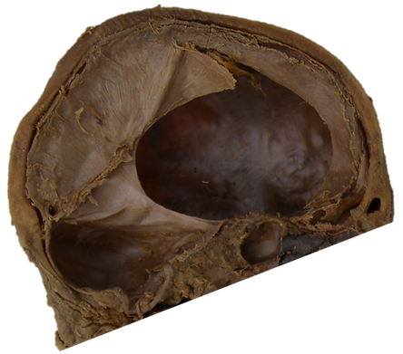
At the posterior margins of the tentorium, two lateral sinuses leave the confluence of sinuses, the transverse sinuses. Follow the canal in the lateral direction and notice how it winds down the sigmoid sinus to the internal jugular vein that drain all the blood and CSF from the brain.